By Dr Brenda Baker BDs (Hons.) (syd.) Msc. Restorative Dentistry (London) consultant at Southern Cross Dental and Modern Dental Pacific.
Dental implants offer an effective and predictable way to replace teeth. Function, aesthetics and phonetics are regained. The long-term success clinically and aesthetically depends upon the appreciation and management of the peri-implant tissues.
Factors contributing to predictable osseointegration include: (Tagliareni JM., Clarkson E., 2015):
- Atraumatic surgery.
- Placement of the implant with initial stability.
- Immobility of the implant.
It is the responsibility of the dentist to address the patient’s concerns, fears and expectations and outcome of treatment. Managing expectations is critical to achieve realistic outcomes for both the dentist and the patient. “Patients should be informed of the spectrum of potential complications and maintenance issues that can occur with implant-borne prostheses and informed of the biological consequences and associated future costs (Lewis MB. and Klineberg I., 2011)”.
The pre-operative assessment should include:
- Chief presenting complaint.
- Medical history and risk assessment.
- Dental history.
- Intraoral evaluation – hard and soft tissues, jaw relationships, parafunction.
- Diagnostic casts and photographs.
- Radiographic examination – presurgical imaging allows clinicians to determine quality and quantity of available bone.
“The clinical examination should take account of oral hygiene and periodontal status, evaluation of the remaining dentition, malocclusion or crowded or rotated teeth, incisal opening, vertical dimension, interarch space, TMJ examination and width of available bone at the edentulous site” (Barber P. and Seckinger RJ., 2000).
At the time of the examination the charting should include (Klein M., 2001):
- Status of remaining teeth.
- Mobility.
- Furcations.
- Periodontal probing depths.
- Keratinised tissue.
- Interarch space.
- Distance between the teeth.
- Ridge width nSupra-eruption.
- Tilted teeth.
- Occlusal/incisal plane.
- Smile line.
- Appearance of soft tissue in the smile.
- Number of teeth in a wide smile.
- Aesthetics of existing teeth.
- Presence of any infection.
The predictability of the aesthetic outcome of an implant restoration depends on: (Jivrai S. et al., 2013):
- Patient selection and smile line .
- Tooth position.
- Root position of the adjacent teeth.
- Biotype of the periodontium and tooth shape.
- Bony anatomy of the implant site.
Researchers have studied when implants have been placed adjacent to natural teeth and implants placed next to each other (Tarnow DP. et al., 1992). It was observed that it was challenging to obtain an interdental papilla when the implants were placed less than 3 mm apart. Implants should be placed at least 3 mm apart to minimise the loss of vertical bone height. Achieving or creating soft tissue interdental papilla in the aesthetic zone is not a predictable outcome. The shape of clinical crowns which have longer contact areas will improve the appearance and may be considered an option.
The following two cases have successfully achieved functional and aesthetic results in the replacement of anterior teeth which were extracted due to trauma.
Case I
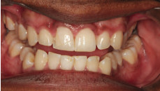
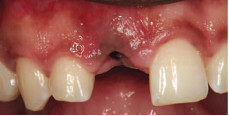
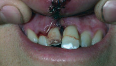
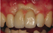
The patient presented to the surgery after a football accident when he was knocked in the mouth (Fig.1). The trauma and pain caused the patient to present to the local hospital for pain relief.
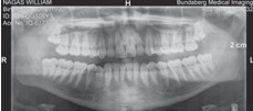
Upon attendance at the surgery, the patient complained of severe pain and sensitivity to cold and air. On examination there was a crack in the tooth which had occurred at an angle and was inclined towards the root of the tooth. An OPG was taken (Fig.2).
The patient was advised that the tooth was unable to be saved.
As a result of potential bone infection, the patient was advised to seek prompt treatment as, if the tooth was left for too long, bone could be lost. A warning was given that some type of graft may be required.
Various treatment options and costings were presented to the patient:
- Bridge.
- Partial upper chrome.
- Implant.
After the treatment plan discussions, the patient opted to have the implant. The relevant literature from the ADA was given to the patient.
Procedures:
1. Implant placement
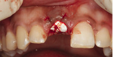
Scandonest 3% was employed with 1.8ml buccal infiltration and Articaine 4% 1:100,000 adrenaline 1.8ml buccal infiltration and 1.8ml palatal infiltration. An alginate impression was taken to make an Essix retainer with a tooth. Elevators and forceps were used to remove 11. The bone was prepared for placement of an implant into 11 socket. The implant placed was IA43-12d-13 4.3 mm X 13 mm TRI-NEX CO-AXIS 12 degree angled (LOT 084D04d2n02) which was torqued to 50Ncm. A 3.5 X 3.0 mm WIDE healing abutment was placed. Bio-Oss .25g graft was placed around the implant and into the socket to keep the ideal gum shape. The Bio-Oss and top of the implant were covered with Cytoplast™ GBR-200 membrane to help keep the bone graft material intact. The socket area was sutured to keep the membrane in place (Fig.3).
An Essix retainer was provided for the patient later in the day.
Panadol and Nurofen were given post operatively and the patient was advised that the bone graft material would have a white sandy texture.
The patient was asked to avoid eating anything hard or crunchy for the next week or so.
2. Stabilisation
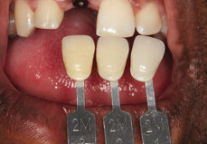
This was scheduled for 2 months after the implant had been placed. An impression of the implant was taken without the need for local anaesthetic. A shade was taken -2M1 2M2 2M3. (Fig.4a and 4b).
The healing abutment was removed and a 3.5 mm impression coping was placed. The radiograph showed that all looked good.
Upper and lower polyvinylsiloxane impressions were taken using AFFINIS® heavy and light body.
The occlusal registration was taken using Blue Velvet for 30 seconds. An explanation of the difficulty of colour matching a single anterior tooth was necessary to ensure the patient understood early reporting back to the dentist if the aesthetics were not excellent. The temporary crown was to be fabricated at the laboratory. The patient understood the need for the provisional restoration to allow ideal tissue recontouring.
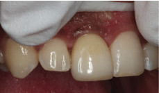
3. Insertion of provisional crown
Local anaesthetic was used Articaine 4% 1:100,000 Adrenaline 1 mls buccal infiltration palatal infiltration quadrant 1. The healing cap was removed and a temporary crown placed on 11 to 32Ncm (Fig. 5).
The access hole was filled using silicone tape. Metal primer Surpass® 2 and 3 (self-etching bonding system) and CLEARFIL MAJESTY™ Flow A2 G-aenial™ P-A2. The occlusion was checked, adjusted and polished. Post-operative Panadol was advised.
4. Definitive crown placement – step 1
A new impression was taken two months later. The gum had remodelled nicely around the gingival margin. Once the temporary crown was removed, the top of the implant was cleaned and the surrounding gum was irrigated with Savacol. A 3.5 mm impression coping was used to take an impression with AFFINIS® heavy and light body silicone. The temporary crown was reinserted and the access hole filled with silicone tape, surpass and A2 flowable and polished.
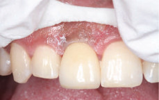
5. Final crown placement – step 2
The shape of the crown was evaluated at the beginning of the visit. The patient and the dentist were happy with the shape and shade of the crown. The new implant abutment crown was screwed in to 35Ncm and the crosspin tightened (Fig.6). At a subsequent appointment, the opposing 42 was adjusted by 0.5 mm. Tooth 42 was anesthetised with Articaine 4% 1:100,000 Adrenalin 1.8 ml by buccal and lingual infiltration in the 42 area. The 42 was checked and adjusted to ensure that the 42 was not hitting hard on the 11 crown.
The 42 was sealed using GLUMA® and CLEARFIL™ SE BOND. It was critical to ensure that no deleterious forces were placed over the implant. The screw was tightened on the 11 implant. “Overload caused by improper prosthesis design or parafunctional habits is considered to be one of the primary causes of late-stage implant failure” (Balshi TJ., 1996).
Case II
The patient presented after an accident when he was hit in the mouth with a generator handle. He had smashed his front teeth. The upper lip was swollen and had been heavily sutured. (Fig.7).
The dentist suspected root fracture of the 21 probably half way up the root. The 11 was considered to have a dubious prognosis. It was deemed that exodontia was probable. The lower anterior teeth had been chipped and required restorations and on one of the teeth the pulp was exposed.
Upon radiographic examination, the 21 had a fracture half way up the root and 11 was deemed to also need exodontia. Initial stabilisation occurred with a removable partial denture. Long-term, the patient was advised to consider implants with two crowns.
Clinical Procedure
Exodontia of 11, 21 with Articaine 4% 1:100,000 Adrenaline .9mls Buccal and palatal infiltration quadrant 1 and 2. 11 and 21 were removed using elevators and forceps. 21 snapped off and had to be f ished out with a size 70 endodontic file. Surgicel was placed in the sockets to help haemostasis and Panadol was given to take home. Impressions were taken for an immediate partial upper denture. The following day, an immediate partial upper denture was issued with the normal post-operative instructions.
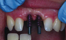
Clinical placement of Implants
Three months later implants were placed. 3.6 cc Lignospan was delivered by infiltration in the 12, 22 area. The incision was made in the gum and pushed back from the bone. A precision drill was used, then a twist drill and then a preparation drill. Implants were inserted into 11 and 21.
IA-LH-35-11.5 3.5mm X 11.5mm TriNex Titanium Implant LOT: 260A02/2 and 13283d2. 3mm healing caps were placed. The implants were torqued 21 to 30Ncm and 11 to 42Ncm. The surgical site was sutured with Vicryl rapide 4/0 suture. The denture was adjusted to fit over the implants.
Postoperatively, the patient was given Panadeine Forte 20 tablets 2 tablets 4 hourly PRN and Amoxil 20 tablets 500mg 1 tablet 3 times a day for a maximum of 7 days. One week later the partial denture was adjusted. IMPRESSION PROCEDURE Articaine 4% 1:100,000 Adrenaline 1.8mls buccal infiltration in quadrant 1 and 2. The healing caps were removed and a shade taken. The impression copings were screwed down and an impression taken with Affinis heavy and light body (Fig.8).
Impression Procedure
Articaine 4% 1:100,000 Adrenaline 1.8mls buccal infiltration in quadrant 1 and 2. The healing caps were removed and a shade taken. The impression copings were screwed down and an impression taken with Affinis heavy and light body (Fig.8).
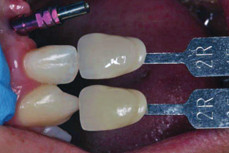
Shade selection was performed at this appointment (Fig.9). The healing abutments were replaced. A lower impression was taken with an occlusal registration.
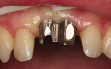
Final issue of Crowns
Articaine 4% 1:100,000 Adrenaline 1.8mls buccal infiltration quadrant 1 and 2. The healing caps were removed and the abutments were screwed into the implants to 32Ncm. Silicone tape was placed over the access hole and the crowns cemented using Fuji cement (Fig.10).
The occlusion was checked to ensure that there was no heavy loads placed on the implant crowns from the other teeth, then the restorations were polished.
The final crowns are shown in situ in Fig.11. Southern Cross Dental would like to thank Dr Lincoln Harris, Bundaberg, Queensland for provision of these clinical cases.
Bibliography
Barber HD, Seckinger RJ. Coordination in the comprehensive diagnosis and treatment of the implant patient: the relationship between the implant surgeon and the restorative doctor. In: Fonseca RJ. Editor. Oral and maxillofacial surgery. St Louis (MO): Saunders Elsevier; 2000. P.14-6.
Balshi TJ. An analysis and Management of Fractured Implants: A Clinical Report International Journal of Oral & Maxillofacial Implants . 1996, Vol. 11 Issue 5, p660-666. 14p
Klein M. Atlas of minor oral surgery. In: Dym H, Ogle O, editors. Evaluation and treatment planning for implants. Philadelphia. WB Saunders; 2001. P.225-30.
Jivraj S., Reshad M, Chee W. Treatment planning of implants in the aesthetic zone: part three. J Implant Pract 2013;6: 32-5.
Lewis, M. B., and I. Klineberg. “Prosthodontic considerations designed to optimize outcomes for single-tooth implants. A review of the literature.” Australian dental journal 56, no. 2 (2011): 181-192.
Tagliareni, Jonathan M., and Earl Clarkson. “Basic Concepts and Techniques of Dental Implants.” Dental clinics of North America 59, no. 2 (2015): 255-264.
Tarnow, Dennis P., Anne W. Magner, and Paul Fletcher. “The effect of the distance from the contact point to the crest of bone on the presence or absence of the interproximal dental papilla.” Journal of periodontology 63, no. 12 (1992): 995-996.